
Knee MRI: a clinical reference pageWhen is an MRI of the knee useful at first contact?
An MRI is useful at first contact when its answer will change what happens next and the clinic receiving the patient can see the images: a knee that is lax after an injury, a knee that will not fully straighten after an injury, a suspected PCL injury, or mechanical symptoms that persist. It is not the first test for a wearing knee, and a report of a degenerate meniscal tear is not a reason to refer.
Written for the clinician seeing the patient first
This page is written for first contact practitioners, advanced practice physiotherapists, MSK physiotherapists and GPs. It is not advice for patients, and it does not replace local pathways or the clinician’s own judgement. Where a statement is Dr Imbuldeniya’s own clinical position rather than published guidance, it says so.
Can the receiving clinic see the images?
An MRI is only worth requesting from first contact if the surgeon who will see the patient can view the images and the report. Orwyn asks this once, as a local setting for the service. Where the answer is no, it writes the referral only, and says why.
Where the scan belongs on day one
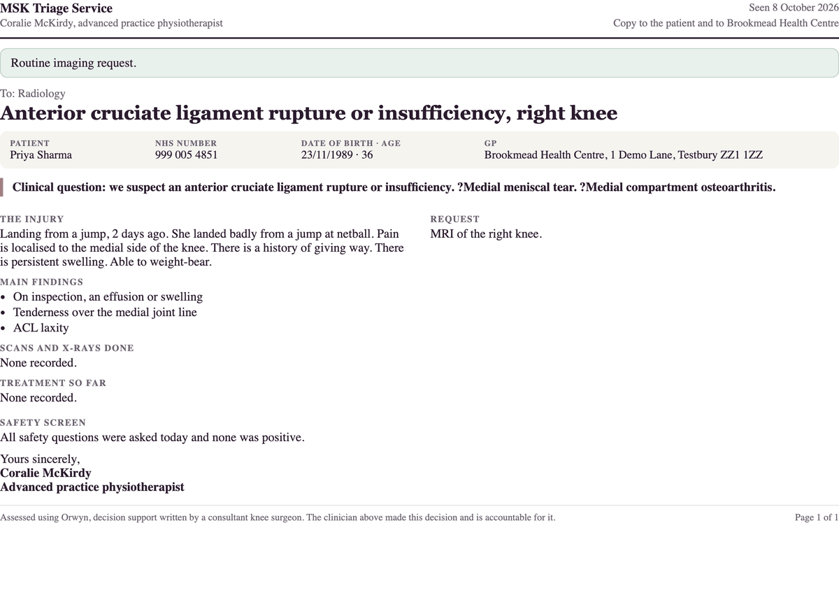
- A knee that is clearly lax after an injury, with the clinician suspicious of a ligament rupture: refer the same day to the acute knee clinic, request the MRI and start physiotherapy, all at once. If the scan is normal, that is acceptable. The BASK and BOSTAA best practice document sets assessment in an acute knee injury clinic within 2 weeks of presentation, with MRI as the imaging of choice.
- Two or more ligaments lax after an injury (ACL, PCL, MCL or LCL; valgus opening counts as MCL laxity) may mean the knee dislocated. Under 4 weeks from the injury, Orwyn sends the patient to A&E (the emergency department) today. At 4 weeks or more, it sends them to the acute knee service, to be seen within 2 weeks. If a foot pulse is weak or different from the other side, it is a limb emergency: call 999.
- A knee that is less certainly lax: request the MRI, start physiotherapy and review within 2 to 6 weeks, when the knee can be examined properly.
- A knee that still will not fully straighten weeks after an injury, in a knee that was good before: urgent referral to the acute knee clinic, to be seen within 1 to 2 weeks, with the MRI requested at the same time.
- A suspected PCL injury: MRI to confirm it, because a posterolateral corner injury can look the same on examination. The report should say whether it is an avulsion, a sprain or a mid-substance tear. An avulsion or a corner injury goes to the acute knee clinic within 2 weeks.
- A second patellar dislocation: urgent referral with the MRI requested. After a first dislocation, the BOA standard reserves MRI for a suspected associated injury, a difficult assessment, planned surgery, or failed non-operative treatment.
- Sharp catching on loading weeks after a twist, in a stable knee that was good before and has never locked, with normal X-rays and physiotherapy not helping: request the MRI and refer to the acute knee clinic at the same time, to be seen within 2 weeks, because the problem is mechanical.
- A suspected loose body (catching that frees itself, a knee that never stays locked): request the MRI and refer to the orthopaedic knee clinic for removal, where the patient would consider an operation.
Where the scan comes later
- A meniscal tear after an injury, in a knee that is not locked, is stable and straightens fully: physiotherapy first, with reassurance, and no MRI on day one. The physiotherapist sends the patient back if the knee is no better at 3 weeks, and the clinician reviews either way. The MRI is requested if pain increases on deep bending, twisting or turning, and the scan does not commit the patient to surgery. The BASK meniscal guideline gives no age criterion, no MRI timeframe and no referral urgency for the injured knee that is not locked.
- Pain at the front of the knee without arthritis: MRI only after 3 to 6 months of physiotherapy.
- A normal examination and normal X-rays: the strength programme first; MRI at about 3 months if the patient is keen or the knee is getting worse.
The wearing knee, and the degenerate tear
For a knee that is wearing, the first test is a weight-bearing X-ray, not an MRI. An MRI is added only when the X-rays are essentially normal and the source of the pain is unclear. The Evidence-Based Interventions guidance does not recommend MRI for a suspected degenerate meniscal tear unless there are mechanical symptoms, and considers it after 3 months of non-operative treatment if symptoms persist.
A patient who arrives with a report of a “degenerate meniscal tear” is being told about early wear, not an injury. Surgery is not offered for a degenerate tear, because the evidence is against it. If treatment is still needed later, the operation that helps is normally a partial or total knee replacement, not keyhole surgery on the tear.
Tears that change the urgency
- A displaced bucket-handle tear: urgent referral to the acute knee clinic, to be seen within days. See the locked knee.
- A bucket-handle tear that is not displaced, a radial tear or a root tear: these can often be repaired and the timing matters. Refer to the acute knee clinic, to be seen within 2 weeks. The patient is referred even if they would prefer to avoid an operation, because the timing matters; the letter records their preference, and they decide with the surgeon.
- New catching after a meniscal repair: the MRI can show a displaced repair or a new tear. It cannot show whether an undisplaced repair has healed, because healing is normally by fibrous tissue, which shows as high signal. High signal at the repair site is not, on its own, a failed repair.
How Orwyn handles this
When the plan includes an MRI, Orwyn writes the request with the clinical question filled in and the findings behind it, and copies the mechanism of injury where it was recorded. Where the receiving clinic cannot see scans ordered by the service, it writes the referral without the scan and says why.
The patient letter explains the scan in one sentence: “An MRI shows the soft structures in the knee: the ligaments, the cartilage and the shock-absorbing pads (menisci), which X-rays cannot.”
Where this came from
- Knee MRI for suspected meniscal tears. Evidence-Based Interventions, Academy of Medical Royal Colleges. Read it here.
- Knee MRI when symptoms are suggestive of osteoarthritis. Evidence-Based Interventions, Academy of Medical Royal Colleges. Read it here.
- Abram SGF, Beard DJ, Price AJ, et al. Arthroscopic meniscal surgery: a national society treatment guideline and consensus statement. Bone Joint J 2019;101-B(6):652-659. Read it here.
- Best practice for management of anterior cruciate ligament (ACL) injuries. British Association for Surgery of the Knee and British Orthopaedic Sports Trauma and Arthroscopy Association, published by the British Orthopaedic Association, 2020. Read it here.
- BOAST: Assessment and management of first time lateral patellar dislocation. British Orthopaedic Association, December 2024. Read it here.
Surgeon-governed decision support for first-contact MSK clinicians.